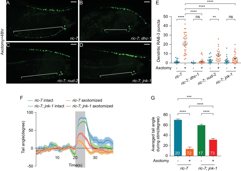
Figure 8. JNK-1 and dynein-mediated transport mediate SV mislocalization to the dendrite and loss of JNK-1 improves behavioral recovery.
(A–D) GFP::RAB-3 localization 48 hr after axotomy in ric-7(n2657) (A), ric-7(n2657); dhc-1(js319) (B), ric-7(n2657); nud-2(ok949) (C) and ric-7(n2657); jnk-1(gk7) (D) animals. Asterisk indicates cell body and bracket indicates dendrite. Scale bars = 10 μm. (E) Quantification of the number of dendritic RAB-3 puncta in intact and axotomized animals 48 hr after axotomy. Mean ± SEM. **p<0.01; ****p<0.0001; ns, not significant. Unpaired t test. (F) Traces of the tail-bending behavior of ric-7(n2657) and ric-7(n2657); jnk-1(gk7) animals with and without axotomy 48 hr after axotomy. The shaded area indicates the 5 s stimulation. Mean ± SEM. (G) Averaged tail angle during stimulation of the animals in (F). Numbers represent the number of animals. Mean and SEM. ***p<0.001; ****p<0.0001. Unpaired t test.


