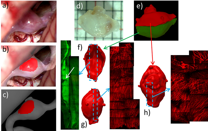
Figure 15:

Identifying and marking aneurysm 3 wall regions from operative video, and MPM imaging of tissue sample: a) operative video frame showing exposed aneurysm, b) superposed video frame and 3D vascular model for image-based marking, c) thin (red) region marked on vascular model, d) photograph of resected sample, e) cutting of tissue sample and alignment of corresponding half tissue models, f-h) MPM imaging of luminal (f) and abluminal (g) side of bottom half of sample, and abluminal side of upper half (h).
