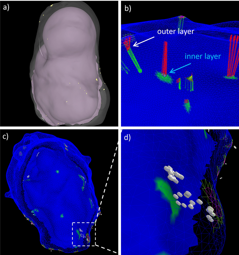
Figure 6:

Projecting micro calcifications: a) inner and outer surfaces of tissue sample with micro-calcifications (small yellow dots), b) distance from calcifications to inner (green arrows) and outer (red arrows) surfaces, c) projection of calcifications to wall layers, and d) detail of projected calcifications (white surfaces) in a small region of the wall.
