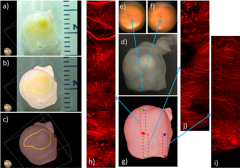
Figure 9:

Identifying and marking aneurysm 1 wall regions from ex-vivo photograph of resected sample, and MPM imaging of tissue sample: a) photograph of resected sample, b) superposed photograph and 3D tissue model for image-based marking, c) “atherosclerotic” region marked on tissue model, d) image of resected tissue sample with ink markings visible under microscopy (e,f) used as reference points to document the location of MPM images, g) marking regions of MPM images on tissue model, h-j) mosaics of MPM image stacks corresponding to the regions marked on tissue model.
