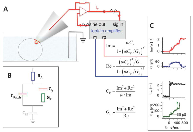
Fig. 4.
Cell attached capacitance measurements of fusion pore dynamics in cells with small vesicles. (A) Cell attached configuration using the lock-in amplifier technique. (B) Minimal equivalent circuit for analysis of fusion pore conductance with components RA, CM, CV, and GP. In this configuration the measured capacitance is that of the patch (CPatch). CM is negligible because it is much larger than CPatch and enters with its reciprocal value. (C) Recording of fusion pore opening from a human neutrophil [11]. GP rises from an initial value as small as 35 pS. CV was calculated from Re and Im as indicated.

