
Figure 3.
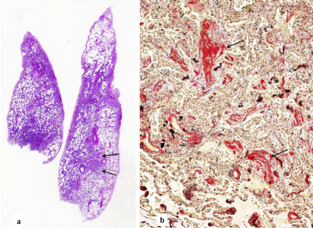
The histology of the biopsied lung. a) Panoramic images of Hematoxylin and Eosin staining lung specimens. On the left side (biopsied from segment 4a of the right lung), diffusely spreading interstitial pneumonia was obvious. On the right side (from the diaphragmatic part of segment 5), areas of relatively distinctly partitioned healthy lung and nodule-shaped intraluminal organization were clear (arrows). b) The nodular area was stained for elastic fibers. The lung structure was indistinct because of interstitial thickening and intraluminal organization (arrows). Magnification: ×100