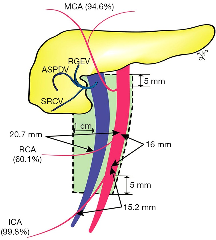
Figure 1.

The boundaries of the D3 area (green area) and the frequency of presence for the ileocolic artery (ICA), right colic artery (RCA), and middle colic artery (MCA). It can be observed the ICA and RCA crossing lengths, and the pooled distance between the ICA to RCA origin distance [Figure reproduced from (20), CC BY license].
