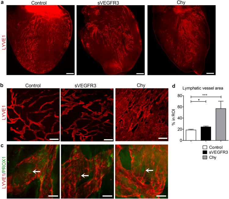
Figure 3.
Cardiac lymphatic vessels are dilated in healthy sVEGFR3 mice and completely loose their organization in Chy mice. (a) Representative images of LYVE1 positive subepicardial cardiac lymphatics in the anterior side of the heart in the healthy control mice, sVEGFR3 mice and Chy mice. (b) Representative images of LYVE1 positive cardiac lymphatic vessels in LVW in the healthy control mice, sVEGFR3 mice and Chy mice. (c) Representative images of PROX1 and LYVE1 double-stainings in LVW in the healthy control mice, sVEGFR3 mice and Chy mice. Arrows indicate PROX1-positive LEC nuclei. (d) Quantification of LYVE1 stainings (n = 11/control mice, n = 17/sVEGFR3 mice and n = 4/Chy mice). Scale bar in (a) is 1,000 µm, in (b) 200 µm and in (c) 50 µm. Values represent mean ± SEM. Statistical analyses were performed using Student’s t-test. *P < 0.05, ***P < 0.001.

