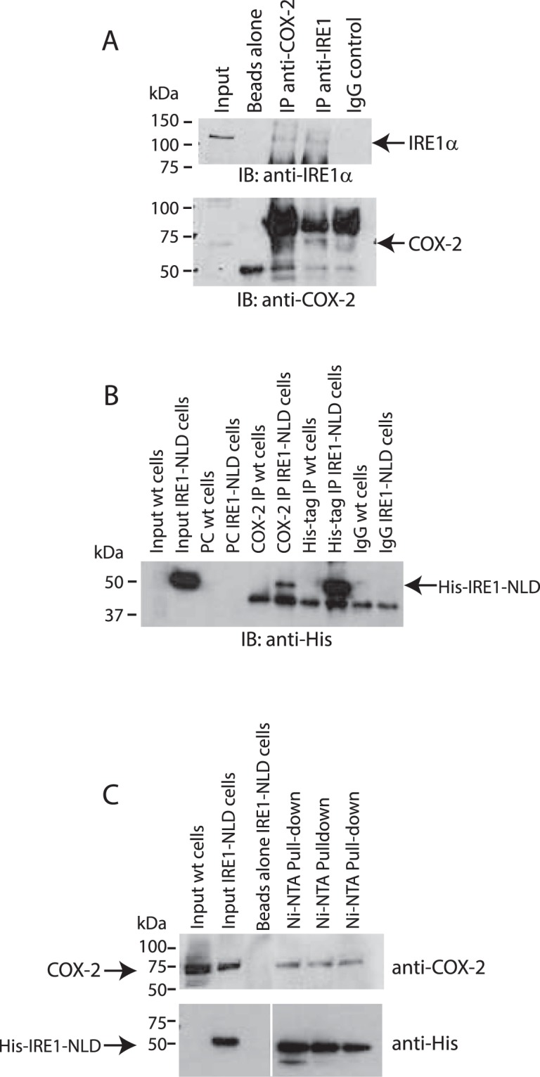
Figure 5.

COX-2 interacts with IRE1α in vivo. (A) Immunoprecipitation (IP) of endogenous IRE1α from HEK293 cells with anti-IRE1α or anti-COX-2 -antibodies. Immunoblot (IB) analysis was carried out with anti-IRE1α or anti-COX-2 antibodies. The location of IRE1α and COX-2 are indicated by the arrows. Immunoprecipitation experiments were performed in triplicate with representative blots shown. (B) His-tagged ER luminal domain of IRE1α (IRE1-NLD) was expressed in COS-1 cells followed by immunoprecipitation with anti-COX-2, anti-His-tag antibodies or IgG. Immunoblot (IB) analysis was carried out with anti-His antibodies. The location of IRE1-NLD is indicated by the arrow. Immunoprecipitation experiments were performed in triplicate with representative blot shown. (C) Pull-down of COX-2 in COS-1 cells expressing His-tagged IRE1-NLD. Upper blot was probed with anti-COX-2 antibodies. The lower blot was probed with anti-His-tag antibodies. Protein samples were separated on the same SDS-PAGE, the middle empty lanes were removed from the lower blot for the sake of clarity. Pull-down assay was performed in triplicate. The images of (A–C) shown are cropped. The full-length gels/blots are shown in Suppl. Figs S12 and S13.
