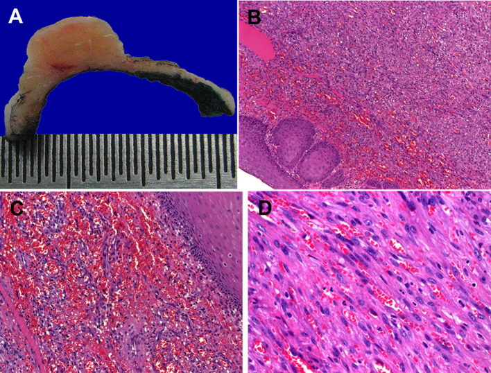
Fig. 2.
Gross and histological appearance of classical KS in the head and neck. a Fleshy cut-surface of the ear lesion shown in Fig. 1a. b Fascicles of uniform spindle cells are covered by reactive non-dysplastic mucosa. c Prominent hemorrhagic changes in the most superficial areas may closely resemble angiosarcoma. d Fascicles of uniform spindle cells entrapping slit-like vascular spaces containing erythrocytes

