
Fig. 16.
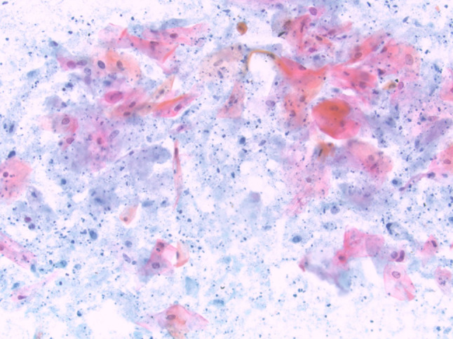
FNA from a cystic squamous cell carcinoma metastatic to the salivary gland showing keratinized squamous cells with only mild atypia present in a background of cystic debris (Papanicolaou stain, magnification × 20)
Official websites use .gov
A
.gov website belongs to an official
government organization in the United States.
Secure .gov websites use HTTPS
A lock (
) or https:// means you've safely
connected to the .gov website. Share sensitive
information only on official, secure websites.
FNA from a cystic squamous cell carcinoma metastatic to the salivary gland showing keratinized squamous cells with only mild atypia present in a background of cystic debris (Papanicolaou stain, magnification × 20)