Fig. 3.

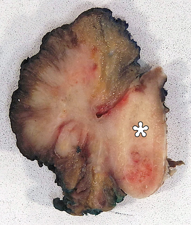
The gross specimen reveals an ill-defined, firm, tan-white mass centered on the floor of mouth with invasion of lingual skeletal muscle and the mandible (asterisk), without distinguishable residual normal sublingual gland

The gross specimen reveals an ill-defined, firm, tan-white mass centered on the floor of mouth with invasion of lingual skeletal muscle and the mandible (asterisk), without distinguishable residual normal sublingual gland