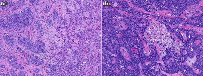
Fig. 4.
Hematoxylin and eosin-stained sections show two distinct areas: a relatively well-differentiated carcinoma with nested, duct-like growth containing cells with high nuclear:cytoplasmic ratios, oval nuclei with coarse chromatin, and variably prominent nucleoli (a, left side); and a more poorly-differentiated carcinoma with solid, sheet-like growth with pleomorphic nuclei and increased mitoses (a, right side). Occasional tumor necrosis was present in areas of solid-to-nested growth (b)

