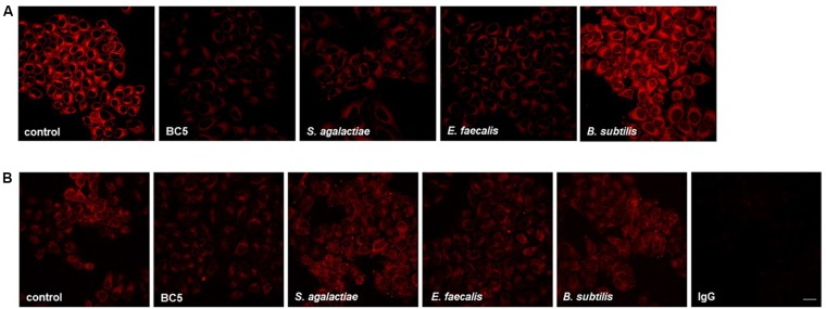
FIGURE 3.
Membrane lipid organization and α5 integrin exposure on HeLa cells incubated with microorganisms. (A) HeLa cells were incubated with L. crispatus BC5, S. agalactiae, E. faecalis, or B. subtilis for 1 h and then stained with NR. (B) HeLa cells were incubated with L. crispatus BC5, S. agalactiae, E. faecalis, B. subtilis for 1 h and then stained for α5 integrin subunit. IgG represents specificity staining control. Representative micrographs are shown. Experiments were repeated at least three times with similar results. Bar: 20 μm.

