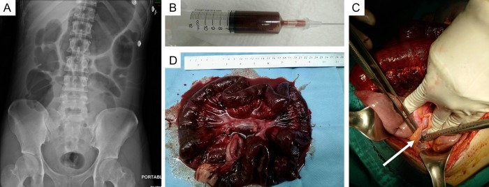
Figure 1:
(A) Abdominal X-ray showed dilated small bowel without free air. (B) Ultrasound-guided paracentesis revealed unclotted blood. (C) One hundred-centimeter segment of necrotic jejunum secondary to adhesion band (arrow) was found at the time of exploratory laparotomy. (D) Resected specimen showed diffuse dark discoloration of the ischemic segment of mid-small intestine.

