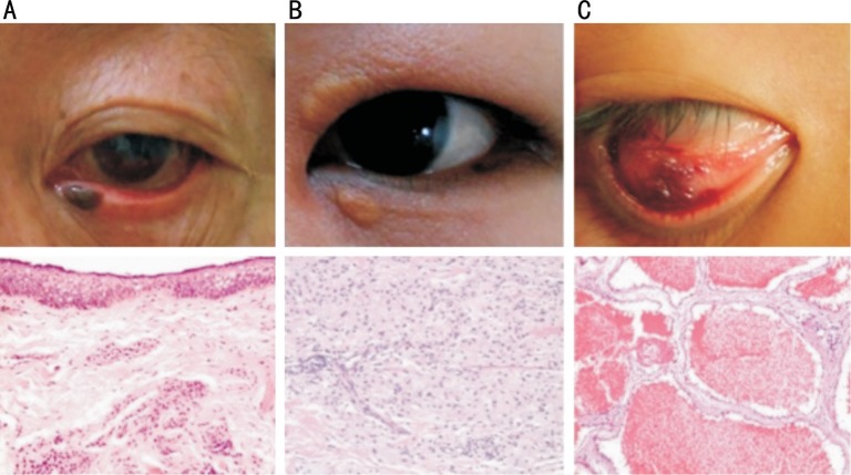
Figure 3. Examples of histopathologic views of miscellaneous tumors.
A: Melanocytic nevus located at the margin of lower eyelid near the inner canthus (H&E 100×); B: Xanthelasma located at the upper and lower eyelids near the inner canthus (H&E 100×); C: Cavernous hemangioma located at the inner side of lower eyelid (H&E 100×).

