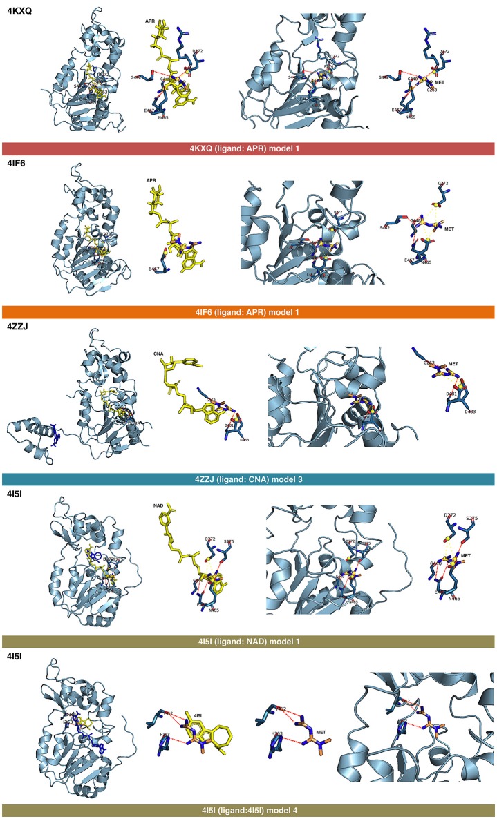
Figure 3.
Self-docking poses under molecular dynamics simulations modeling the metformin binding mode to the APR, CNA, NAD+, and 4I5 binding pockets of SIRT1. Overall structure and views of the interaction between metformin and the APR, CNA, NAD+, and 4I5 binding pockets of SIRT1. The coordinating residues are numbered.

