Figure 3.
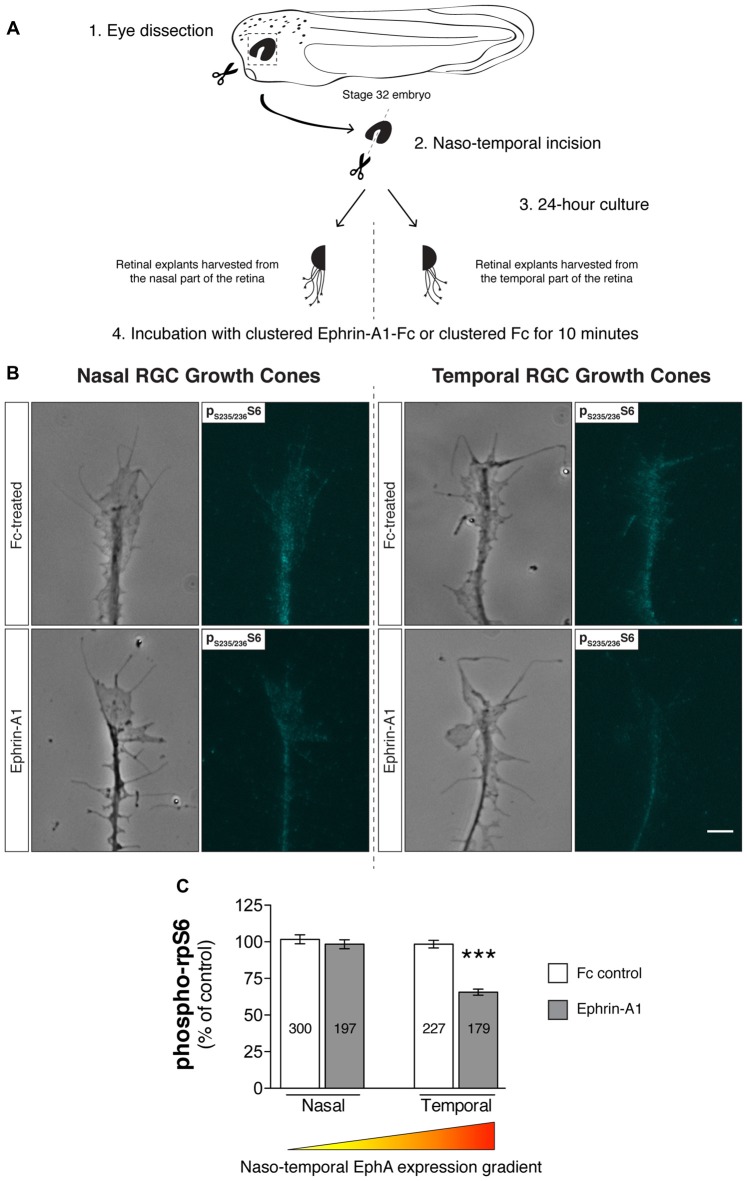
Retinal ganglion cell (RGC) origin dictates distinct growth cone mTOR activation profiles in response to Ephrin-A1. (A–C) Nasal and temporal stage 32 retinal explants grown in vitro for 24 h were stimulated with Ephrin-A1-Fc at a concentration of 5 μg/mL, and stained with an antibody that specifically recognizes ribosomal protein S6 (rpS6) when phosphorylated at Ser-235/236. Representative micrographs of control (clustered Fc)- and Ephrin-A1-treated RGC growth cones are shown (mean ± SEM; n = no. of growth cones analyzed; ***P < 0.0001, one-way ANOVA). Scale bar: 5 μm.

