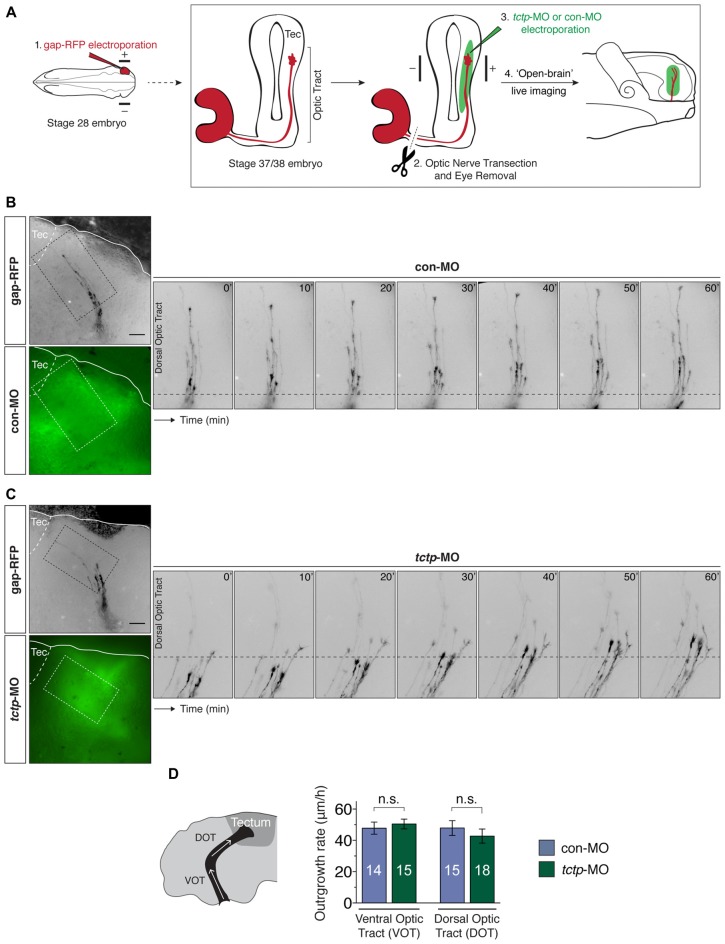
Figure 5.
Local synthesis of Tctp is not required for axon extension in vivo. (A) Gap-RFP was delivered by eye-targeted electroporation to wild-type stage 28 embryos, when the first RGC axons have just exited the eye, and axon extension kinetics were analyzed 21–24 h later (stage 37/38), when most axons are growing up the optic tract towards the optic tectum. Just before imaging, the optic nerve was manually transected, and control morpholino oligonucleotide (con-MO) or tctp-MO was delivered subcellularly by targeted brain electroporation. (B,C) Time-lapse series of severed RGC axons coursing through the optic tract in embryos subcellularly electroporated with con- or tctp-MO. (D) Quantication of axon outgrowth rates through the VOT and DOT in embryos subcellularly electroporated with con- or tctp-MO (mean ± SEM; n = no. of axons analyzed; VOT: P = 0.5956, unpaired t-test; DOT: P = 0.4381, unpaired t-test; 7–9 replicates were analyzed per condition; n.s., not significant, unpaired t-tests). Scale bars: 50 μm.

