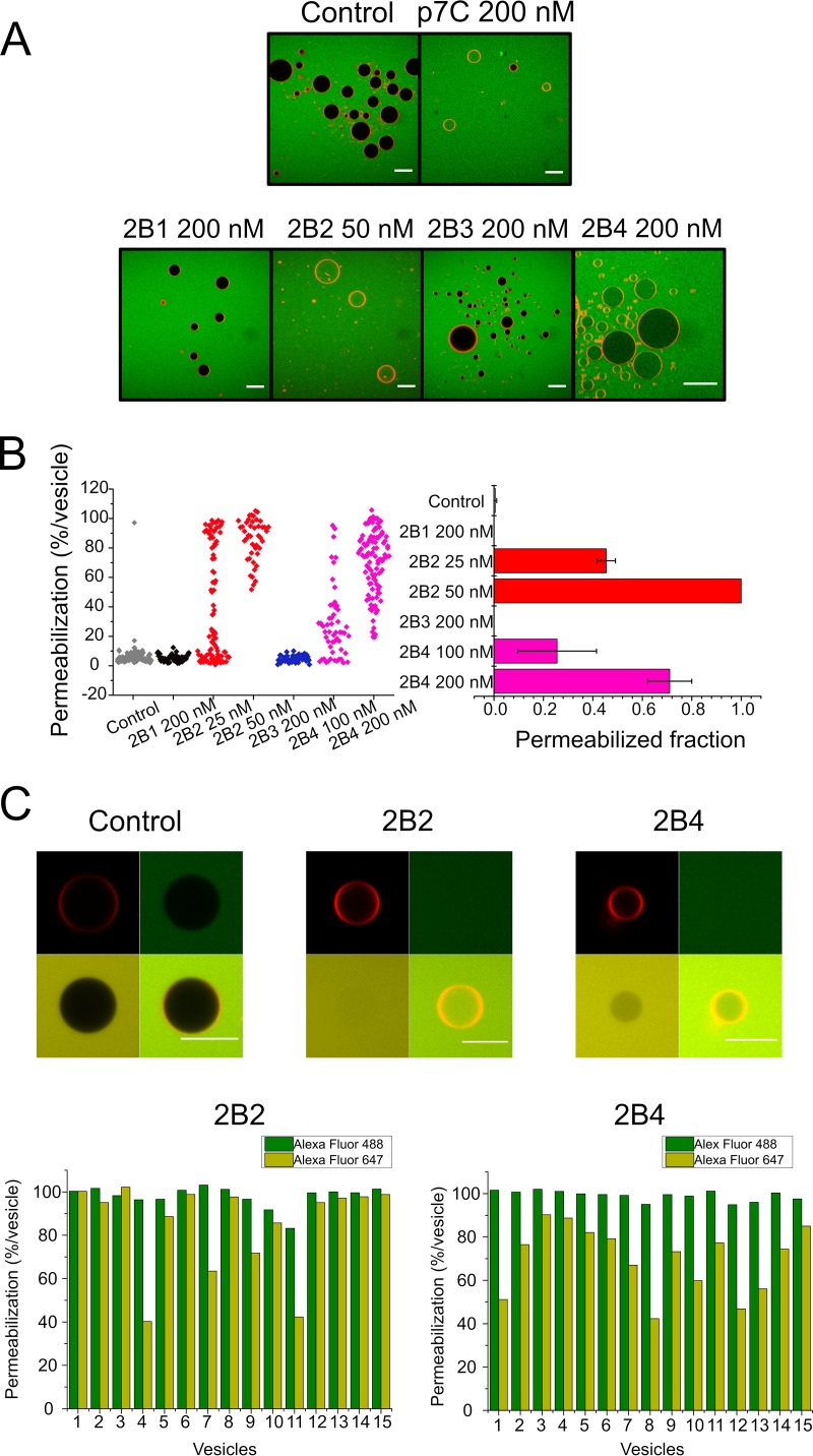
FIG 3.
Single-ER-GUV permeabilization induced by 2B peptides. (A) Micrographs depicting Rho-PE-labeled ER-GUVs (orange circumferences) immersed in a solution containing Alexa Fluor 488 (green background). Samples on top correspond to control untreated ER-GUVs (left) or ER-GUVs treated with the p7C peptide derived from CSFV p7 viroporin (11). Bottom samples correspond to ER-GUVs treated with different 2B peptides at the doses displayed on the panels. Bars correspond to 25 μm in all micrographs. (B) Distribution of ER-GUVs according to their percentage of permeabilization to Alexa Fluor 488 after treatment with the different 2B peptides (left) and mean permeabilization values in the samples (right). Peptides were applied at the doses displayed on the panels. (C) Stability of the membrane permeabilization state induced by 2B2 and 2B4. (Top) ER-GUVs permeabilized to Alexa Fluor 488 after incubation for 2 h with 2B2 or 2B4 (green) were supplemented externally with Alexa Fluor 647 (red) and additionally incubated for 2 h before image processing. The presence of both probes inside vesicles can be observed in the merged images (bottom right panels). The “Control” panel displays an intact vesicle incubated in the absence of peptide. (Bottom) Bars represent the degree of filling of individual ER-GUVs with Alexa Fluor 488 (dark green) and Alexa Fluor 647 (light green) after a 4-h incubation with the 2B peptides.

