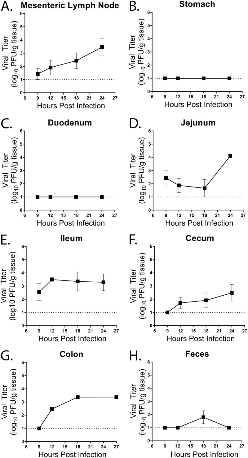
FIG 1.
Kinetics of MNV-1-NR infection in WT mice. WT mice were infected by oral gavage with 3.8 × 10⁵ PFU/animal of neutral-red-labeled MNV-1. Mesenteric lymph nodes (A), stomach (B), duodenum (C), jejunum (D), ileum (E), cecum (F), colon (G), and feces (H) of five mice per time point were harvested at the indicated times in a darkened room using a red photolight. The tissue homogenate was serially diluted and exposed to white light for 30 min. Replicated viral titers in the indicated tissues were assessed via plaque assay. The detection thresholds are indicated by dotted lines. The data are from two independent experiments. The error bars represent standard errors of the mean (SEM).

