
FIG 1.
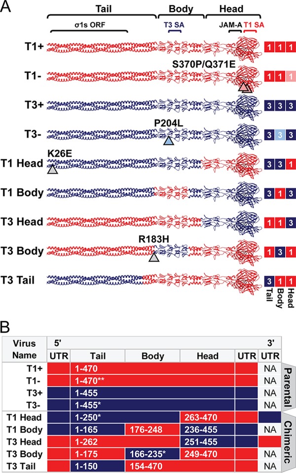
Chimeric σ1-encoding viruses used in this study. (A) Schematic of engineered σ1 proteins. A model of the σ1 trimer (adapted from reference 12) is shown as a ribbon diagram (left) or a simplified box schematic (right). T1 sequences (red; 1) and T3 sequences (blue; 3) are indicated for the tail, body, and head domains. T1 and T3 parental strains bind glycans (+) or contain mutations that abrogate glycan binding (−). Mutations that differ from T1+ and T3+ are indicated by arrowheads and noted. Approximate sites of the overlapping σ1s ORF and receptor binding domains for T1 sialic acid (SA), T3 SA, and JAM-A are annotated. The schematic on the right indicates sequence origin of σ1 domains for each virus (red, T1; blue, T3). (B) T1 (red) or T3 (blue) S1 gene elements are depicted for each virus. The approximate σ1 head, body, and tail domains are noted. Native untranslated regions (UTR) are shown for 5′ and 3′ gene termini. S1 genes of T1 Head and T3 Head viruses contain additional UTR sequences appended to the 3′ termini. Amino acid boundaries are provided for chimeras and numbered by parental strain origin (1 to 470 of T1 and 1 to 455 of T3). Asterisks indicate the number of amino acids that differ from either T1+ or T3+ parental strains in the corresponding domain. NA, not applicable.