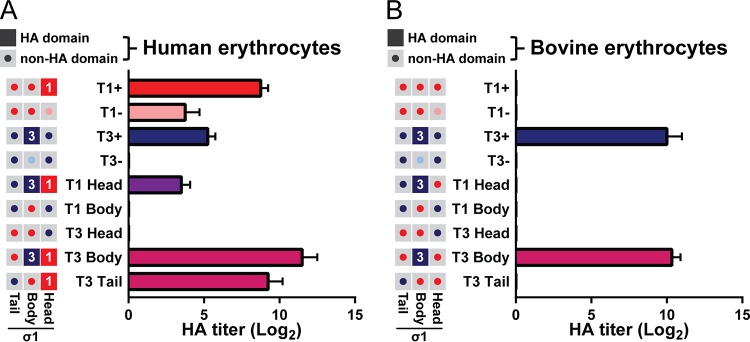
FIG 3.
Chimeric viruses display distinct sialic acid-binding profiles. (A, B) Hemagglutination capacity of parental and σ1-chimeric viruses. Schematics on left indicate the predicted hemagglutination (HA) capacity of σ1 head, body, and tail domains for each virus. Filled boxes (red, T1+; blue, T3+) indicate domains with predicted hemagglutination capacity. Gray boxes with central buttons indicate that the domain is not hypothesized to contribute to hemagglutination (red, T1+; pink, T1−; dark blue, T3+; light blue, T3−). Purified reovirus virions (1011) were serially diluted 2-fold in PBS. Human (A) or bovine (B) erythrocytes were resuspended in PBS at a concentration of 1% (vol/vol). Equal volumes of virus and erythrocyte mixtures were combined and incubated at 4°C for 4 h, and hemagglutination was assessed. Results are expressed as mean log2-transformed HA titers from three independent experiments. Error bars indicate SD.

