FIG 5.
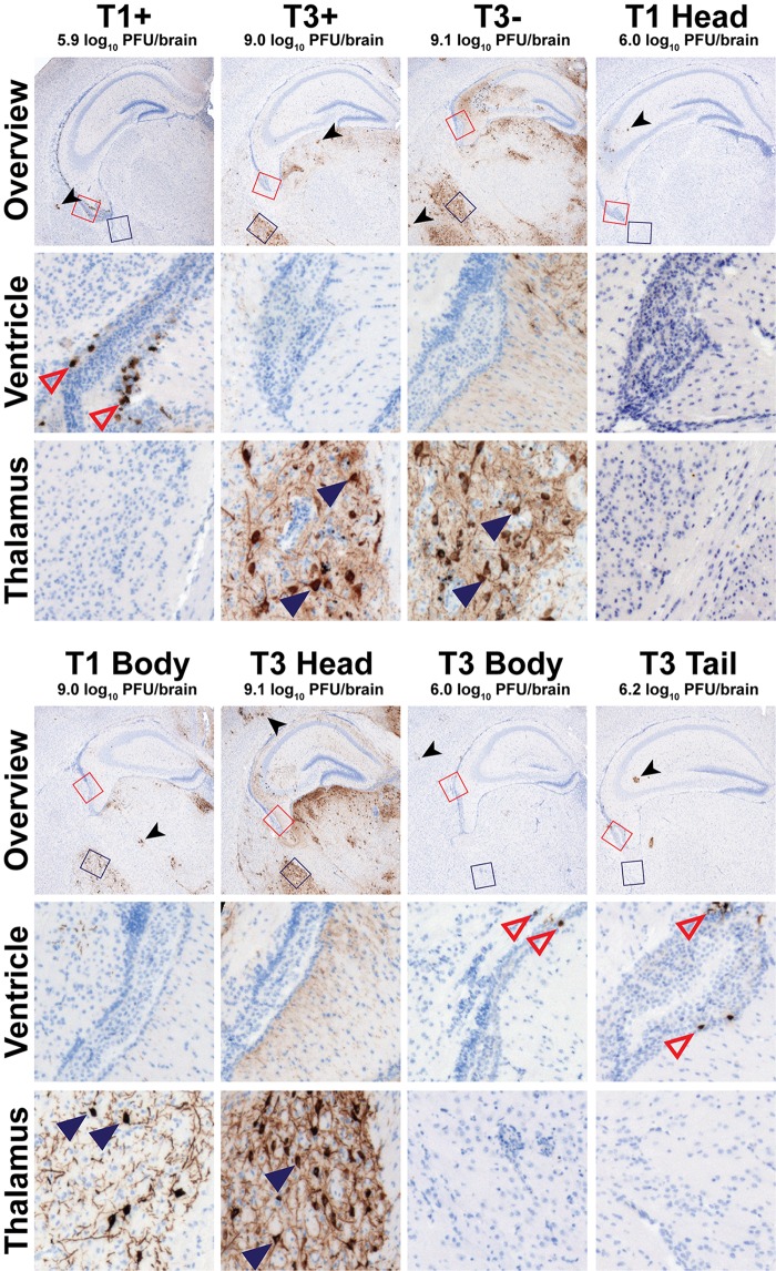
Reovirus neurotropism is dictated by sequences in the σ1 head domain. Newborn C57BL/6 mice were inoculated intracranially with ∼100 PFU of purified virions of the strains shown. Mice were euthanized 8 days postinoculation, and brains were resected and hemisected. Right brain hemispheres were homogenized for determination of viral titer by plaque assay. Left brain hemispheres were fixed in formalin and embedded in paraffin. Coronal sections of the left brain hemisphere were stained with reovirus-specific antiserum and hematoxylin. Low-magnification overview images at the depth of the hippocampus are shown. Regions corresponding to high-magnification insets of the lateral ventricle and lateral thalamus are indicated in the overview micrographs by red or blue boxes, respectively. Representative sections are shown. Viral titers from the paired right brain hemispheres are displayed above the micrographs. Reovirus-infected ependymal cells (open red triangles), neurons (filled blue triangles), and glia (black arrowhead), all identified using morphological criteria, are indicated.

