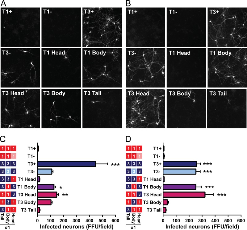
FIG 6.
Infection of cultured cortical neurons is primarily dependent on sequences in the T3 σ1 head domain. Cultured primary rat cortical neurons were treated with a vehicle control (A, C) or 40 mU/ml of neuraminidase (B, D), inoculated with reovirus at an MOI of 500 PFU/cell, fixed at 24 h postinoculation, and stained with reovirus-specific antiserum, an antibody to detect Tuj1, and DAPI. (A, B) Representative micrographs of reovirus-infected neurons (white staining) are shown. (C, D) Infected neurons were enumerated using indirect immunofluorescence. Results are expressed as mean numbers of infected neurons per field of view for six images per well in quadruplicate wells for five independent experiments. Error bars indicate SEM. Values that differ significantly from T1+ by one-way ANOVA and Dunnett's test are indicated (*, P < 0.05; **, P < 0.01; ***, P < 0.001).

