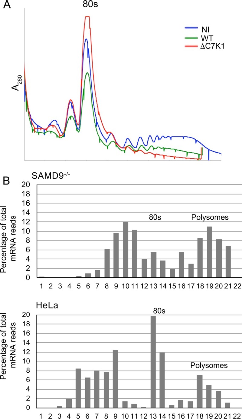
FIG 5.
Localization of viral mRNA on sucrose gradients. (A) Polyribosome profiles. HeLa cells were not infected (NI) or were infected with WT or ΔC7K1 virus for 6 h, after which their cytosols were centrifuged on sucrose gradients. The gradients were collected from the top while we continuously recorded the absorbance (A260) to monitor ribosomes and polyribosomes. The 80S ribosome position is indicated. (B) Distribution of D13 mRNA. HeLa and SAMD9−/− cells were infected with ΔC7K1, and the cytosols were centrifuged on sucrose gradients as described for panel A. RNA was extracted from the resulting fractions and reversed transcribed, and D13 mRNA was quantified using ddPCR. Fraction 1 is the top of the gradient.

