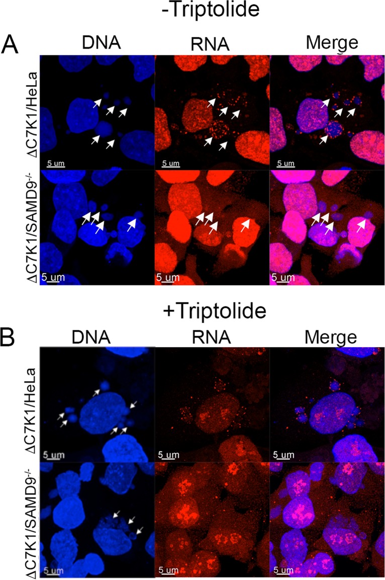
FIG 6.

Localization of EU-labeled nascent RNA. (A) HeLa and SAMD9−/− cells were grown on coverslips, infected with ΔC7K1, and incubated with 1 mM EU from 5 to 6 h after infection. Cells were washed, fixed, and permeabilized, and EU-labeled RNA was detected by conjugating Alexa Fluor 568 azide using click chemistry. DNA was detected by staining with DAPI. Arrows pointing to the positions of the viral factories visualized with DAPI are shown in all panels. Magnification is indicated by size bars. (B) HeLa and SAMD9−/− cells were infected with ΔC7K1, as described for panel A, except that triptolide was added at 4 h and 1 mM EU at 5 h. At 6 h, the cells were fixed and treated as described for panel A. Arrows pointing to viral factories visualized with DAPI are shown only in the DNA panels.
