
FIG 7.
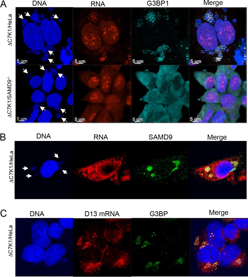
Association of viral RNA with the G3BP stress granule marker and SAMD9. (A) Colocalization of EU-labeled cytoplasmic RNA and G3BP1 at viral factories. HeLa and SAMD9−/− cells were infected with ΔC7K1. At 4 h after infection, cells were treated with 1 µM triptolide for 1 h and then 1 mM EU in the continued presence of triptolide for an additional 1 h. EU-labeled RNA was detected as described in the legend to Fig. 6A, and G3BP was visualized with a rabbit polyclonal antibody followed by a fluorescent secondary antibody. Arrows point to DAPI-labeled DNA factories. Magnification is indicated by size bars. (B) Colocalization of EU-labeled RNA and SAMD9. HeLa cells were infected and analyzed as described for panel A, except that the antibody was to SAMD9 instead of G3BP1. (C) Colocalization of viral D13 mRNA with G3BP1. HeLa cells were infected as described for panel A except for the absence of triptolide. Following fixation and permeabilization, D13 mRNA was detected by FISH using probes to D13 mRNA. G3BP1 was detected with specific antibody.