
Figure 4.
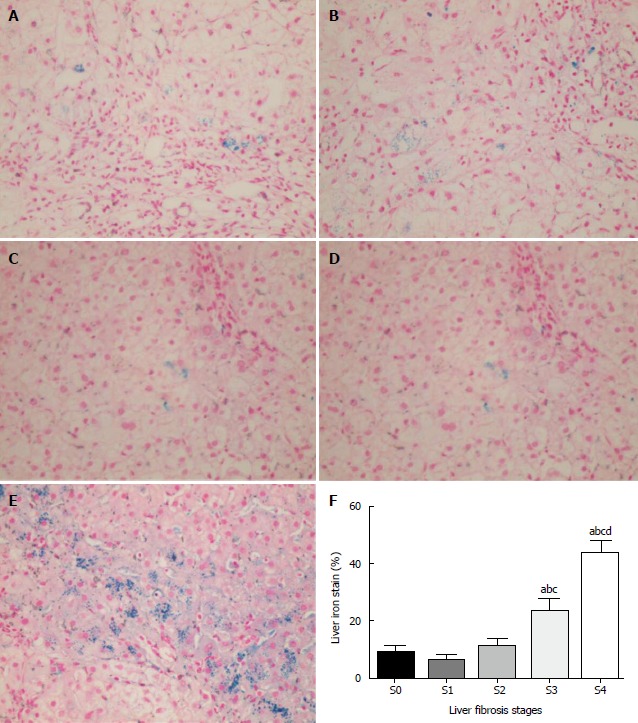
Iron deposition in liver tissues with fibrosis of different stages. Perls’ staining of iron appears as red granular particles in liver cells (× 400 magnification.) A-E: Different stages of liver fibrosis, respectively. Liver fibrosis was staged using the METAVIR scoring system, which consists of five stages: S0 (no fibrosis, n = 5), S1 (portal fibrosis without septa, n = 8), S2 (portal fibrosis with rare septa, n = 4), S3 (portal fibrosis with many septa, n = 7), and S4 (cirrhosis, n = 5). Markedly increased iron deposition was observed in the severe liver fibrosis (S3) and cirrhosis (S4) groups, but not in groups S0-S2. F: Significantly higher average iron retention (mean ± SEM) with severe fibrosis (S3: 23.7%) and cirrhosis (S4: 43.6%) compared to that with no or mild fibrosis (S0: 5.2%, S1: 7.9%; S2: 8.5%). A statistically significant difference in iron staining was observed among patients with severe fibrosis and cirrhosis (P < 0.05). aP < 0.05 vs S0; bP < 0.05 vs S1; cP < 0.05 vs S2; dP < 0.05 vs S3.