FIGURE 1.

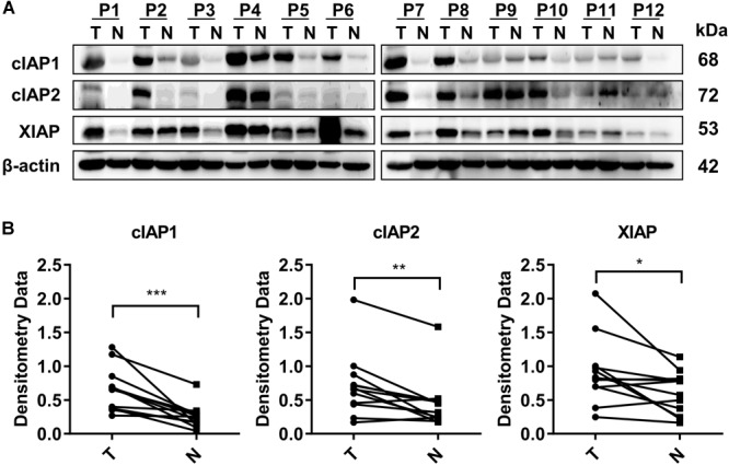
Proteins associated with the inhibitor of apoptosis protein (IAP) gene were highly expressed in HCC tissue. (A) The protein levels of cIAP1, cIAP2, and XIAP in 12 pairs of tissue samples of HCC were measured by Western blot. (B) The bands were semi-quantified by densitometry and normalized to β-actin. P, HCC patient; T, HCC tumor tissue; N, normal adjacent liver tissue; kDa, kilodalton. ∗p < 0.05, ∗∗p < 0.01, ∗∗∗p < 0.001, NS, not significant, by two-tailed pair t-test.
