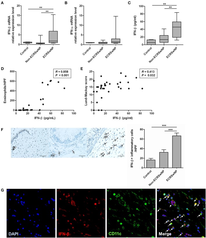
Figure 1.
Expression of type I IFN in human sinonasal tissue. (A,B) IFN-β mRNA and protein levels are enhanced in tissue homogenates from eosinophilic CRS with NP (ECRSwNP) patients. (C) On the other hand, there are no differences in the IFN-α family mRNA expression between control, non-eosinophilic CRSwNP (non-ECRSwNP), and ECRSwNP groups. (D,E) IFN-β level positively correlated with the number of eosinophils in the sinonasal tissue and Lund-Mackay CT score in 31 patients with CRSwNP. (F) In addition, IFN-β+ cells (indicated by arrows) are more frequently detected in the lamina propria of ECRSwNP tissues compared with the controls and non-ECRSwNP (magnification, ×400; scale bar, 50 μm). Horizontal bars indicate the medians. **P < 0.01; ***P < 0.001, repeated measurement ANOVA with Tukey's multiple comparison test (A–C,F) and Spearman correlation test (D,E). (G) In immunofluorescence assay for NP tissue, IFN-β (red fluorescence) was frequently detected in CD11c cells (green fluorescence). Nuclei were counterstained with 4',6-diamidino-2-phenylindole (DAPI; blue fluorescence). In the merged image, cells indicated by arrows show co-localization of IFN-β and CD11c cells. The immunofluorescence results are representative of 5 different subjects (Magnification, ×400).

