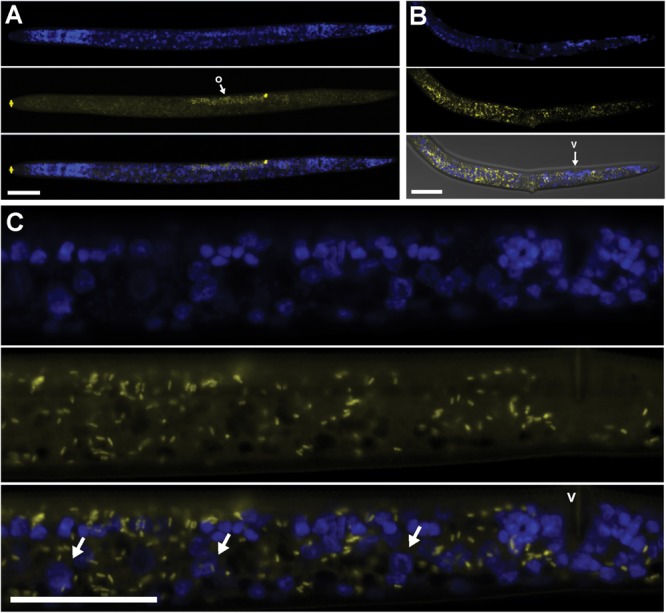
FIGURE 3.

Localization of Cardinium cPpe in whole body and ovaries of P. penetrans by FISH and confocal microscopy. (A–C) Split panels showing nematode nuclei (blue DAPI stain) alone (top), cPpe (yellow FISH probe) alone (middle), and combined images (bottom). (A) Whole female with o = ovaries, and yellow autofluorescence of lip and spermathecal regions. (B) Female with combined brightfield DIC image (bottom), v = vulva. (C) High magnification of (A) showing ovaries and developing oocytes (arrows), v = vulva. Scale bars = 20 μm.
