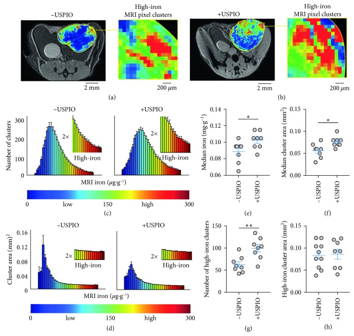
Figure 1.
Spatial quantification of endogenous and nanoparticle-enhanced iron deposits with MRI in vivo. T2-weighted MRI and iron concentration overlay images of (a) control (−USPIO) and (b) iron nanoparticle-injected (+USPIO) tumors. Expansion shows high-iron pixel contrast in clustered areas. (c) Number (#) of clusters and (d) area of the pixel clusters in control (−USPIO) and nanoparticle-injected (+USPIO) tumors as a function of iron concentration. Distributions are from whole cross-sectional regions of interest (ROI) areas of tumors measuring approximately 1 cm3 (mean ± SEM shown, n=8 tumors/group). MRI iron concentration range at bottom corresponds to values in iron images above. Control (−USPIO) and nanoparticle-injected (+USPIO) (e) median iron concentrations and (f) pixel cluster sizes. (g) Number (#) of high-iron pixel clusters and (h) size of the high-iron clusters from localized computer vision analysis (mean ± SEM shown, n=8 tumors/group, n.s. p > 0.05, ∗p < 0.05, ∗∗∗p < 0.001, two-tailed unpaired Student's t-test). Scale bars are shown for all images.

