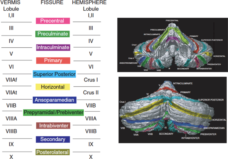
Figure 1.
The cerebellum is organized into three major lobes that can be further sub-divided into 13 distinct lobules, defined by sulcal landmarks, and into the medial vermis and lateral hemispheres. The anterior lobe is divided into lobules I-V, the posterior lobe is divided into lobules VI-IX, and the flocculonodular lobe consists of lobule X. The defining sulcal boundaries are the precentral, preculminate, intraculminate, primary, superior posterior, horizontal, ansoparamedian, prebiventer, intrabiventer, secondary, and posterolateral fissures. Reprinted from Schmahmann, J. D., Doyon, J., Petrides, M., Evans, A. C., & Toga, A. W. (2000). MRI atlas of the human cerebellum. San Diego: Academic Press.

