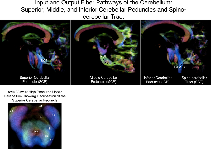
Figure 2.
Input and output fiber pathways of the cerebellum, shown on a high-resolution diffusion-weighted image. Lower panel: Axial slice at the high pons/upper cerebellum showing the decussation of the superior cerebellar peduncle (crossing red fibers marked 23’). Numbering is from Naidich, T. P., Duvernoy, H. M., Delman, B. N., Sorenson, A. G., Kollias, S. S., & Haacke, E. M. (2009). Duvernoy’s atlas of the human brain stem and cerebellum. Vienna: Springer-Verlag/Wien. 16 = corticospinal tract; 21 = medial lemniscus; 23 = superior cerebellar peduncle; 23’ = decussation of the superior cerebellar peduncle; 38 = middle cerebellar peduncle; 41 = ventral pontine decussation; 42 = dorsal pontine decussation; 47 = lateral lemniscus.

