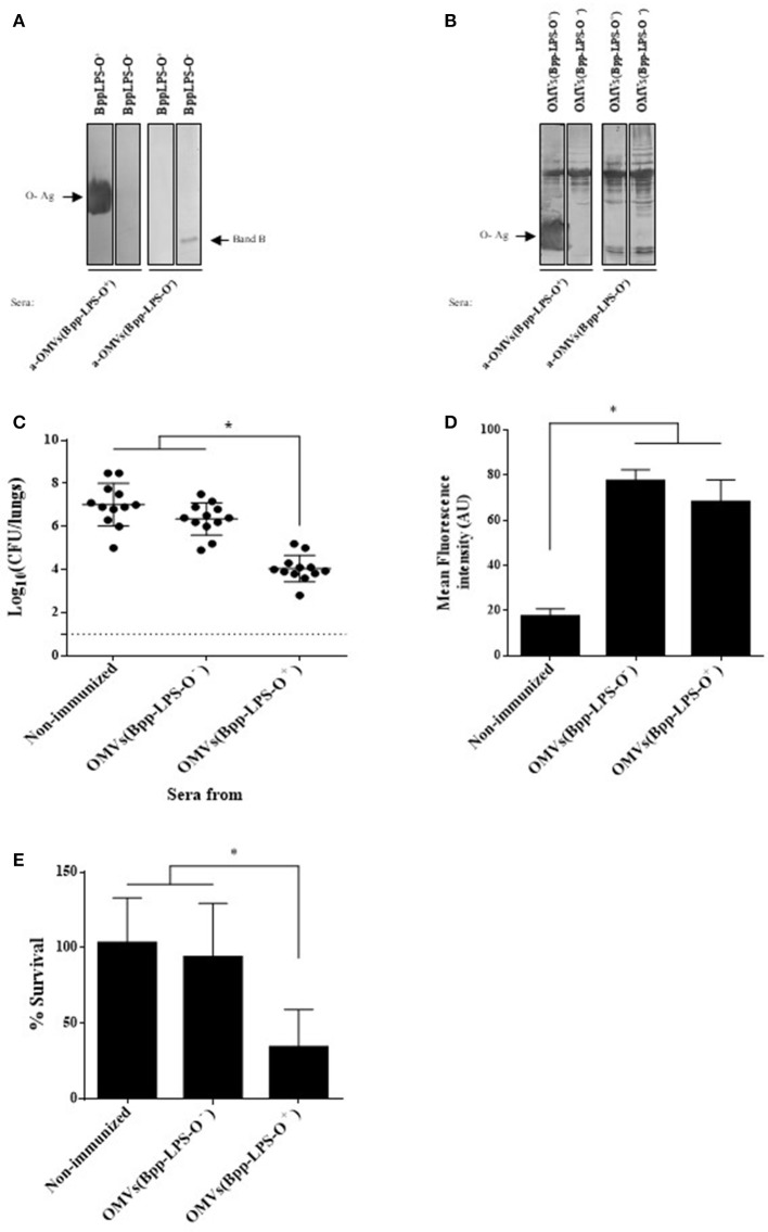
Figure 3.
Humoral response induced by purified BppLPS-O+ and OMVs(Bpp-LPS-O+). (A) Immunoblotting of purified B. parapertussis LPS separated by 15% (w/v) SDS-PAGE probed with the polyclonal antiserum obtained from mice immunized with OMVs(Bpp-LPS-O+) and OMVs(Bpp-LPS-O−). The arrows indicate the locations of the O antigen (O-Ag) and the Band B that were recognized by the antiserum used in the subsequent immunoblotting indicated below the gels. These sera are designated according to the immunogen— OMVs(Bpp-LPS-O+) and OMVs(Bpp-LPS-O−)—used to raise the immune response in the donor mice. (B) Immunoblotting of OMVs(Bpp-LPS-O+) and OMVs(Bpp-LPS-O−) samples separated by 12.5% (w/v) SDS-PAGE and probed with the polyclonal antiserum obtained from mice immunized with OMVs(Bpp-LPS-O+) and OMVs(Bpp-LPS-O−). The arrows indicate the locations of the O antigen (O-Ag) and the Band B that were recognized by the antiserum used in the immunoblotting indicated below the gels. These sera are designated according to the immunogen—OMVs(Bpp-LPS-O+) and OMVs(Bpp-LPS-O−)—used to raise the immune response in the donor mice. (C) Effect of passive immunization with sera collected from OMVs(Bpp-LPS-O+) - or OMVs(Bpp-LPS-O−) -immunized mice. Pooled sera from either OMVs(Bpp-LPS-O+) - or OMVs(Bpp-LPS-O−) -immunized mice were tested by transfer to naive female mice (n = 12). Pooled sera from non-immunized mice transferred to naive female mice were used as a negative control of protection. Twenty-four h after the transfer, the mice were infected with B. parapertussis AR729 as the challenge bacterium (5 × 10⁷ CFUs in 40 μL). In the figure, the number of bacteria recovered from the mouse lungs, expressed as the log10 of the means ± SEM (error bars) of the CFUs per lungs, is plotted on the ordinate for the non-immune and the two immune sera indicated on the abscissa, with the latter being designated according to the immunogen—OMVs(Bpp-LPS-O−) and OMVs(Bpp-LPS-O+) —used to raise the immune response in the donor mice. The asterisk (*) indicates a significant difference vs. the naïve sera (p ≤ 0.05). (D) Opsonophagocytic activiy of the immune sera. The mean fluorescence intensity in arbitrary units (AU) associated with macrophages previously incubated with GFP-expressing B. parapertussis AR729 is plotted on the ordinate for each of the sera used for opsonization of those bacteria on the abscissa, indicated as non-immune or immune, with the latter being designated according to the immunogen—OMVs(Bpp-LPS-O−) and OMVs(Bpp-LPS-O+)—used to raise the immune response in the donor mice. The asterisk (*) denotes p ≤ 0.0001 vs. the intensity obtained after exposure of the bacteria to the non-immunized serm. (E) Serum-complement–mediated killing. Sera from OMVs(Bpp-LPS-O+)—or OMVs(Bpp-LPS-O−)-immunized mice were incubated with B. parapertussis AR729 (500 CFU), and 1 h later the bacteria were plated to determine viability. In the figure, the percent survival is plotted on the ordinate for the non-immune and the two immune sera indicated on the abscissa, with the latter being designated according to the immunogen—OMVs(Bpp-LPS-O−) and OMVs(Bpp-LPS-O+)—used to raise the immune response in the donor mice. The asterisk (*) indicates a significant difference vs. the naïve sera (p ≤ 0.05).

