
FIGURE 1.
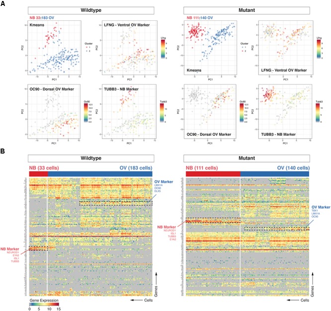
Loss of Chd7 shifts the distribution of cells the E10.5 otocyst toward those with neuroblast identity. Principal component analyses (PCA) (A) and hierarchical clustering (B) on Chd7+/+ and Chd7Gt/+ E10.5 otic derived cells distinguishes otic epithelia cells from putative neuroblast cells and reveals a relative increase in the proportion of Chd7Gt/+ cells expressing pro-neural genes compared to wild type. Cells projected onto first two components are color-coded based on k-means cluster and expression levels of three representative markers (Lfng = ventral otic epithelium, Oc90 = dorsal otic epithelium, Tubb3 = delaminated neuroblasts). This was confirmed by increases in pro-neural (Neurod1, Tubb3) vs. pro-epithelial (Oc90) gene expression amongst Chd7 heterozygous cells.