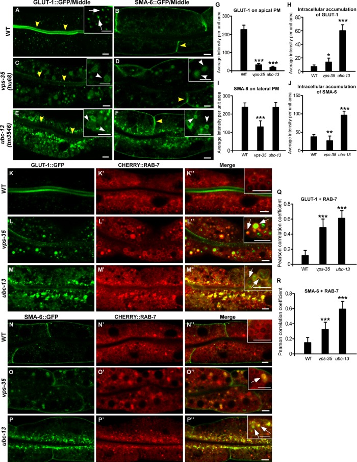
FIGURE 4:
ubc-13 affects recycling of the retromer-dependent cargoes GLUT-1 and SMA-6. (A–F) Confocal fluorescence images of the intestine in wild type (WT; A, B), vps-35 (C, D), and ubc-13 (E, F) expressing GLUT-1::GFP (A, C, E) or SMA-6::GFP (B, D, F). Yellow arrowheads indicate GFP labeling on apical or lateral membranes, white arrows designate GLUT-1::GFP puncta in the cytosol of wild-type worms, and white arrowheads indicate vesicular structures labeled by GLUT-1::GFP or SMA-6::GFP in the cytosol of vps-35 and ubc-13 mutants. (G–J) Quantification of the average fluorescence intensity of GLUT-1::GFP (G, H) and SMA-6::GFP (I, J) per unit area in different genetic backgrounds. (K–P″) Confocal fluorescence images of the intestine in wild-type (WT; K–K″, N–N″), vps-35 (L–L″, O–O″), and ubc-13 (M–M″, P–P″) animals coexpressing CHERRY::RAB-7 and GLUT-1::GFP (K–M″) or SMA-6::GFP (N–P″). Arrows indicate colocalization of GFP and CHERRY. (Q, R) Colocalization of CHERRY::RAB-7 and GLUT-1::GFP (Q) or SMA-6::GFP (R) was quantified in the indicated strains. In G–J and Q and R, at least eight animals were quantified in each strain, and data are shown as mean ± SD. One-way ANOVA with Tukey’s post hoc test was performed to compare all the other data sets with wild type. *, P < 0.05; **, P < 0.001; ***, P < 0.0001. Scale bars: 5 μm.

