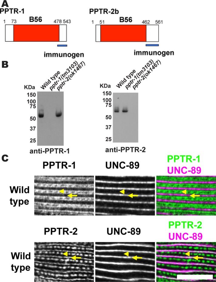
FIGURE 4:
Immunolocalization of PPTR-1 and PPTR-2 to sarcomeres of body-wall muscle. (A) Schematic representation of PPTR-1 and PPTR-2 polypeptides, each having a single domain, the B56 domain (shown in red). Blue bars indicate the unique C-termini used as immunogens to generate rabbit antibodies. (B) Western blots demonstrate that antibodies to PPTR-1 or PPTR-2 detect expected-size proteins from the wild type but not from the respective deletion alleles. The numbers on the left side of each blot show the position of molecular weight markers in kDa. (C) By immunofluorescence microscopy, anti–PPTR-1 localizes to sarcomeric I-bands, and anti–PPTR-2 localizes to sarcomeric dense bodies and M-lines. Portions of a single body-wall muscle cell are shown in each row, costained with antibodies to UNC-89, which localizes to M-lines. Arrowheads mark the positions of dense bodies, and arrows mark the positions of M-lines. In the two-color image at bottom right, white indicates colocalization of PPTR-2 with UNC-89 at M-lines. Scale bar, 10 μm.

