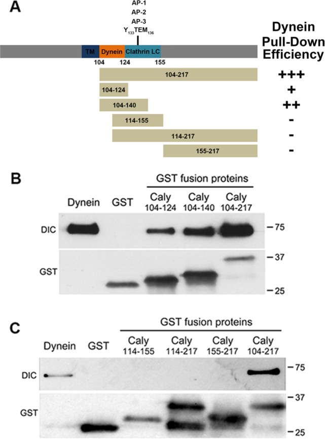
FIGURE 2:
Mapping of dynein binding domain in Caly. (A) Stick figure diagrams the domain organization of the Caly protein, and the segments of Caly fused to GST for pull-down studies. Plus/minus (+/-) signs indicate effectiveness of respective GST fusion protein in pulling down purified bovine brain dynein. (B, C) Immunoblots of pull-down experiments probed with antibodies to DIC and GST. The “Dynein” lane in each blot was loaded with 5 μg of purified dynein. The remaining lanes were loaded with resin-bound material eluted following incubation of GST Caly fusion proteins with the purified dynein complex (100 μg protein). (B) The GST fusion containing juxtamembrane residues 104–124 was the shortest Caly fusion able to pull down dynein. (C) GST-Caly 114–155 was ineffective at pulling down dynein, suggesting that the relevant binding domain lies between residues 104 and 114.

