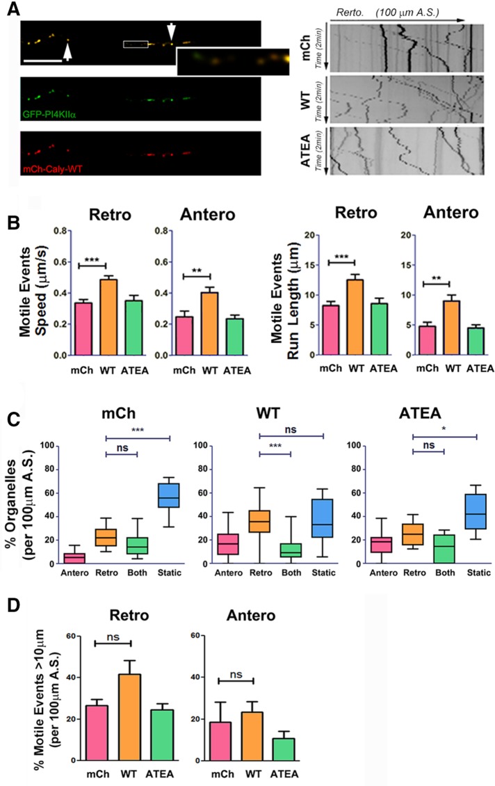
FIGURE 8:
Caly-WT but not Caly-ATEA increases the motility of PI4KIIα-labeled organelles. (A) Left panels, colocalization of GFP-PI4KIIα (green) and mCh-Caly-WT (red). Arrows point to overlapping puncta, and enlarged box show some examples of nonoverlapping puncta. Right panels, representative kymographs of PI4KIIα-labeled organelles in axons transfected with mCherry (mCh), mCh-Caly-WT (Caly-WT), and mCh-Caly-ATEA (Caly-ATEA). (B) Average speeds and run lengths of both anterograde and retrograde motile events are higher in Caly-WT axons, whereas values detected in Caly-ATEA axons do not differ from control. (C) Box-and-whisker plots show that retrogradely moving PI4KIIα-labeled organelles are more common in Caly-WT axons relative to static and bidirectional organelles. (D) No difference in retrograde and anterograde motile events over >10 μm were detected. Data shown in bar graphs (mean ± SEM) and box-and-whisker plots (mean ± 95% CI) reflect results obtained in three independent experiments, with at least five axons per group in each experiment; *p < 0.05, **p < 0.01, ***p < 0.001; A.S., axon segment; bar = 20 μm.

