FIGURE 4:
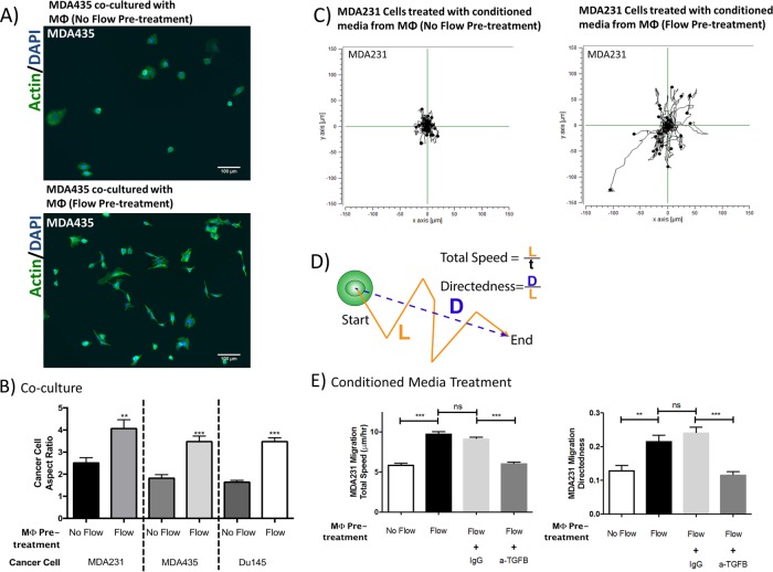
IF enhances the ability of macrophages to promote cancer cell migration. Murine bone marrow–derived macrophages were pretreated with interstitial flow (3 μm/s for 48 h) and their ability to influence cancer cell migration and morphology were assessed (see also Supplemental Figure S6). (A) Representative fluorescent images (green = actin, blue = DAPI) showing that MDA435 cancer cells cocultured with macrophages pretreated with flow (bottom) were more protrusive than cancer cells cocultured with macrophages that were not pretreated with flow (top). (B) Quantification of cancer cell morphology showing that cancer cells cocultured with macrophages pretreated with flow have higher aspect ratio than ones cocultured with control macrophages that were not treated with flow. (C) Representative migration trajectories of MDA231 cancer cells treated with conditioned media collected from macrophages pretreated with flow (right) and of cancer cells treated with conditioned media from control macrophages (left). (D) Definition of cell migration dynamics. L = total migration distance; D = net displacement; t = time. Directedness is a measure of persistence. (E) MDA231 cancer cells treated with conditioned media from interstitial flow-primed macrophages show a higher migration total speed (left) and directedness (right) than cells treated with conditioned media from control macrophages. Inhibition of TGFβ (anti-TGFβ neutralizing antibody, 10 μg/ml) in the conditioned media reduced this increase in migration speed and directedness. Bars represent mean ± SEM of data from 45–100 cells (n = 3).

