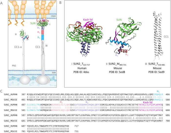
FIGURE 1:
Structures of various fragments of SUN2 in a trimeric or monomeric state in the NE. (A) Schematic representation of the standing model of SUN2 organization in the NE. The in vitro crystal structures available from various studies are shown in their respective positions, and all other regions for which no experimental structural information is available are shown with schematic cartoon representations. Note that the organization of α1 and α2 in the trimer, as well as CC1-α in the monomer, is unknown. (B) Available structures of SUN2 fragments: (i) structure of Human Sun2522–717 trimer bound to three KASH peptides, (ii) structure of Mouse SUN2483–731 monomer, and (iii) structure of coiled coil (CC1) of Mouse SUN2420–481. Each structural fragment is represented by its residue range. SUN2_M is used for the monomeric state of each fragment and SUN2_T for a trimeric state. (C) Amino acid sequences of Human and Mouse SUN2. Structural domains on Human and Mouse Sun2 are shown with similar colors for comparison (α3 in purple, α2 in pink, α1 in cyan, Sun domain in green, and the KASH lid in magenta).

