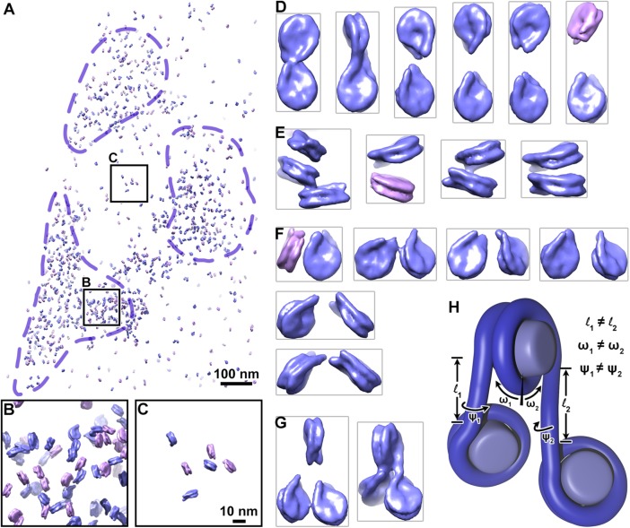
FIGURE 3:
Chromatin is irregular at the oligonucleosome level in situ. (A) Model of short-linker (magenta) and long-linker (blue) nucleosomes remapped according to their positions and orientations in the nucleus. Dashed purple lines indicate approximate boundaries of heterochromatin. (B, C) Fourfold enlargements of the heterochromatin and euchromatin positions boxed in A. (D–G) Examples of (D) dinucleosomes connected by linker DNA. (E) face-to-face packed nucleosomes, (F) dinucleosomes not connected by linker DNA but likely to be in sequence with a third nucleosome that was missed by our analysis, and (G) trinucleosomes connected by linker DNA. For clarity, adjacent remapped nucleosomes were cropped out. (H) Schematic of a trinucleosome, showing histone octamers (light blue) and DNA (dark blue). The lengths (l1, l2), angles relative to the dyad axis (ω1, ω2), and rotation around the linker-DNA axes (ψ1, ψ2) are uncorrelated.

