FIGURE 5:
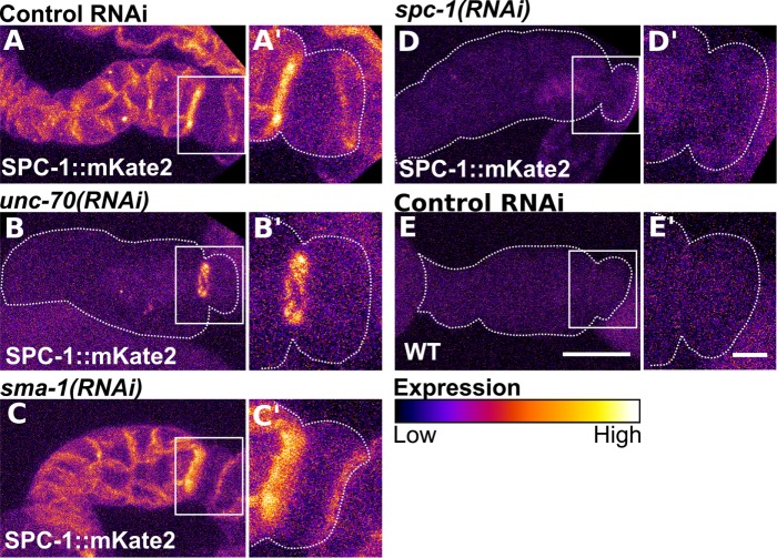
The localization of SPC-1/alpha-spectrin requires UNC-70/β-spectrin in the spermathecal bag and both β-spectrin isoforms in the valve. Confocal maximum projections of excised and fixed spermathecae from animals expressing SPC-1/α with a CRISPR generated mKate2 tag at the endogenous locus (SPC-1::mKate2) treated with control RNAi (A), RNAi against unc-70 (B), sma-1 (C), and spc-1 (D). Note the loss of SPC-1/α at spermathecal cell boundaries with depletion of UNC-70/β (B) but retention of SPC-1/α in a band connecting the spermathecal bag and SP-UT valve (B′). SPC-1/α localization is not changed with SMA-1/βH depletion (C–C′). (E) Confocal maximum projections of an excised and fixed spermatheca from a WT, unlabeled, animal appears similar to the SPC-1::mKate2 line treated with RNAi against spc-1 in D. Inserts in A′–E′ are a magnified view of the valve indicated by a white box in A–E. Images are colored to highlight differences in fluorescence intensity. Scale bars, 20 μm, 5 in inserts.

