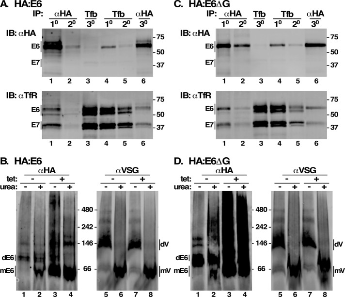
FIGURE 5:
Misfolding of E6 reporters. (A, C) Cell extracts were prepared from HA:E6- or HA:E6ΔG-expressing cell lines without silencing of native TfR. Aliquots (107 cell equivalents) were sequentially immuno/affinity precipitated in two sets [1° and 2° αHA, 3° Tf-beads (Tfb), lanes 1–3; and 1° and 2° Tfb, 3° αHA, lanes 4–6). Pull downs were subjected to simultaneous immunoblotting with mAb anti-HA and rabbit anti-TfR using Licor IR-fluorescent secondary antibodies with different emission wavelengths. The anti-HA (top) and anti-TfR (bottom) signals were digitally separated for presentation. Mobilities of E6/E7 subunits and molecular mass markers (kDa) are indicated. (B, D) Cell extracts were prepared with and without RNAi silencing (tet +/-) and fractionated by Blue Native gel electrophoresis (106 cell equivalents/lane) with and without denaturation (urea +/-). Gels were transblotted and probed simultaneously with mAb anti-HA and rabbit anti-VSG and specific Licor secondary reagents. Anti-HA and anti-VSG signals were digitally separated for presentation. Mobilities of E6 dimers (dE6), E6 monomers (mE6), VSG dimers (dVSG), VSG monomers (mVSG), and molecular mass markers (kDa) are indicated.

