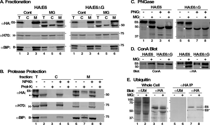
FIGURE 7:
Characterization of MG132 protected species. All analyses were performed after RNAi silencing (24 h) of endogenous TfR. All MG132 treatments were for 2 h at 25 μM. (A) HA:E6- and HA:E6ΔG-expressing cells were incubated without (Cont) or with MG132 (MG). The cells were hypotonically lysed and total (T), cytoplasmic (C), and membrane (M) fractions were prepared. All fractions (107 cell equivalents/lane) were analyzed by immunoblotting with anti-HA (αHA), anti-Hsp70 (αH70, cytoplasmic marker), or anti-BiP (αBiP, ER marker). (B) Cell fractions prepared from MG132-treated HA:E6 cells were treated with proteinase K (Prot-K) as indicated in the absence or presence of NP40. Samples (107 cell equivalents/lane) were immunoblotted with anti-HA, anti-Hsp70, and anti-BiP. (C–E) HA:E6- or HA:E6ΔG-expressing cells were incubated without (-) or with (+) MG132 treatment. (C) Cells were solubilized under denaturing conditions and each was split into two equal fractions (107 cell equivalents). One set was mock-treated (-) and the other digested (+) with PNGase F (PNG). Samples were analyzed by immunoblotting with anti-HA. (D) Lysates were immunoprecipitated with anti-TfR antibodies (107 cell equivalents/precipitate) covalently cross-linked to Protein A sepharose. One set (right) was immunoblotted with anti-HA (αHA) and the other blotted with biotinylated ConA (left). Mobilities of E6 and molecular mass markers (kDa) are indicated on the left. White strip indicates digital reordering of lanes after image processing. Stars in A–D indicate mobility of MG132-protected E6 or fully deglycosylated species. (E) Total extracts of HA:E6-expressing cells were prepared in SDS sample buffer, and lysates for immunoprecipitation were prepared identically to GPI-PLC-lysates in Figure 8 to minimize deubiquitinating activities. Total cell extracts (lanes 1–4) and anti-HA immunoprecipitates (lanes 5–8) were immunoblotted (107 cell equivalents/lane) sequentially with anti-ubiquitin and anti-HA on the same membrane and the individual signals were processed prior to digital separation for presentation.

