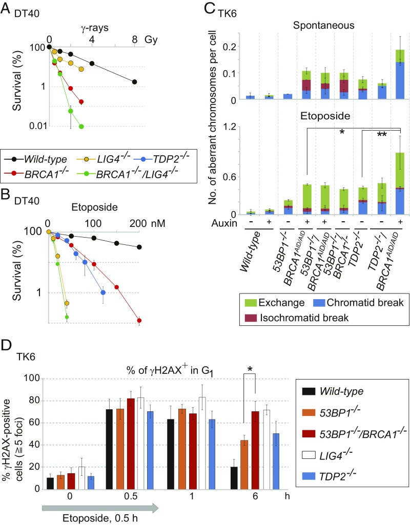
Fig. 2.
Function of BRCA1 is dependent on the canonical NHEJ pathway and independent of TDP2 in the repair of etoposide-induced DNA damage. (A and B) Cellular sensitivity of chicken DT40 cells to γ-rays (A) and etoposide (TOP2 poison) (B) was measured by clonogenic cell-survival analysis. Error bars show SD calculated from three independent experiments. (C) Number of spontaneously arising mitotic chromosome breaks (Upper) and etoposide-induced chromosome breaks (Lower). BRCA1AID/AID cells were treated with auxin (250 μM) (+) or without auxin (−) for 9 h. The cells were treated with etoposide (100 nM) for 9 h (Lower). Cells were incubated with colcemid for the last 3 h. Error bars represent SD of three independent experiments. At least 50 metaphase cells per experiment were examined for the chromosome analysis. The single and double asterisks (Lower) indicate P < 0.03 and P < 0.02, respectively, calculated by Student’s t test. (D) DSB-repair kinetics of TK6 cells in G1 phase following 0.5-h pulse exposure to etoposide (10 nM). Cells were released to fresh medium after the pulse exposure. DSB repair was evaluated by counting the number of γH2AX foci at 0.5, 1, and 6 h after the initiation of the pulse exposure. The percentage of cells with at least five foci per nucleus is shown on the y axis. Error bars were plotted for SD from three independent experiments. The asterisk indicates P < 0.01, calculated by Student’s t test. More than 100 G1 cells (cyclin-A–negative) were analyzed for each experiment. The average numbers of γH2AX foci per cell are shown in SI Appendix, Fig. S8.

