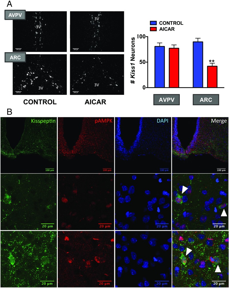
Fig. 3.
Activation of AMPK suppresses Kiss1 expression in the arcuate nucleus. In the Upper panels (A), dark-field photomicrographs showing Kiss1 mRNA expression (white clusters of silver grains) in representative sections of the AVPV and the ARC in the hypothalamus of pubertal female rats (PND33) chronically treated with vehicle or AICAR are presented. In addition, we display data from the quantification of Kiss1 expression in the above experimental groups. **P < 0.01 vs. corresponding control group and nucleus (ANOVA followed by Student–Newman–Keuls multiple range test) (n = 4–5 animals per group). 3V, third ventricle. In the Lower panels (B), confocal images are shown of the expression of kisspeptin and pAMPK in the ARC of pubertal female rats (PND36). In the Top row, low magnification images are shown for Kisspeptin (Left), pAMPK (Central Left), and DAPI (nuclear staining; Central Right); the image on the Right corresponds to the final merge of the three signals. Two different examples at higher magnification are shown in the additional two rows below. Colocalization is denoted by white arrowheads.

