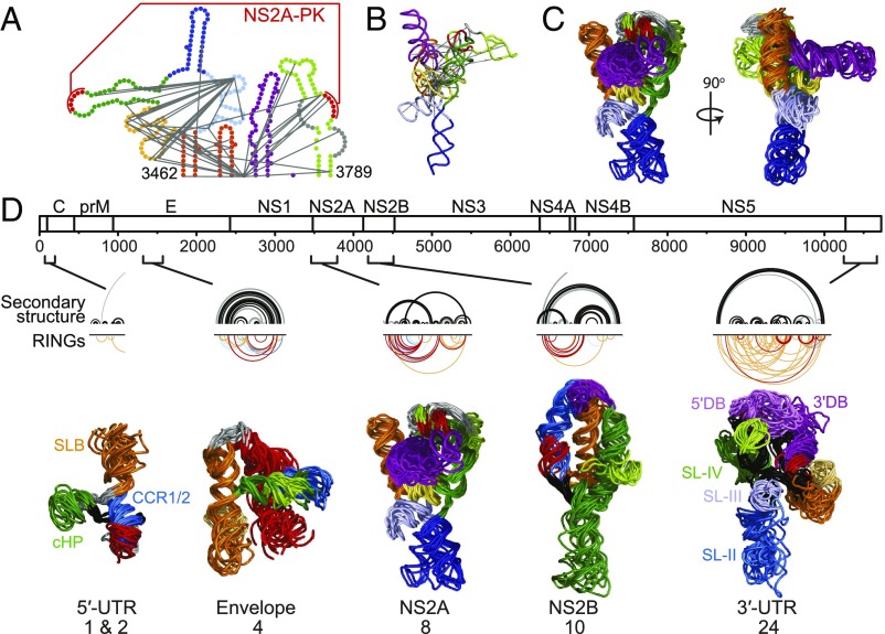
Fig. 6.
Higher-order structures throughout the DENV2 genome. (A) Secondary structure of the NS2A element. RINGs used as tertiary structure constraints in the DMD simulation are depicted by gray lines. (B) Medoid of DMD models of the NS2A element. (C) The 10 lowest aligned free-energy DMD models of the NS2A element. (D) From top to bottom, the DENV2 RNA genome, the secondary structure (black arcs) and tertiary RINGS (inverted colored arcs) of elements with tertiary structures, and tertiary structure models. RINGs reporting tertiary interactions are color-coded by correlation coefficient as in Fig. 2, and 3D folds are color-coded by secondary structure (SI Appendix, Figs. S11–S14). Previously identified elements in the 5′- and 3′-UTRs (7–9) are labeled. For each element, the 10 lowest free-energy models from the largest cluster are shown.

